tree in bud opacities treatment
After chemotherapy was started the patient showed prompt clinical improvement. TIB opacities typically show branching configurations from secondary pulmonary lobules with.

Figure 3 From Tree In Bud Pattern Semantic Scholar
Of these 182 cases were excluded for the following reasons.
. Multiple causes for tree-in-bud TIB opacities have been reported. 4 focal bronchiectasis characterized by a single region of bronchiectasis with. Thus we separated patients with bronchiectasis and TIB opacities into three other patterns.
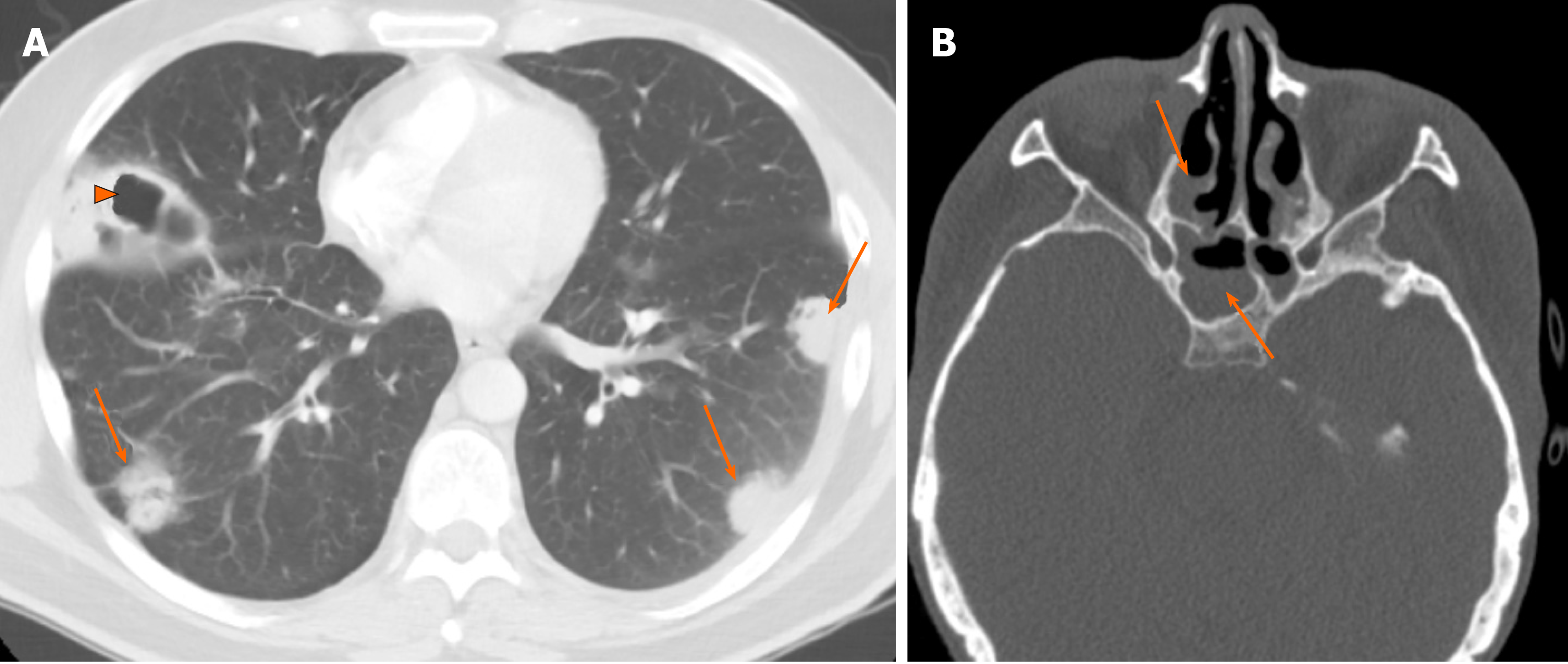
The tree-in-bud lesions were caused by arterial embolization of primary neoplastic cells from an osteosarcoma. Tree-in-bud from January 1 2010 to December 31 2010 iden-tifying 599 examinations. The walking distance before discharge 14 days after initiating chemotherapy improved from 60 m to.
Nuclear Medicine 53 years experience. PDF We report a clinical case of mentally challenged young gentleman who was repeatedly hospitalized for respiratory symptoms. The appearance of a tree in bud is depicted by a pattern of bronchial dilatation and filling on a thin-section chestCT.
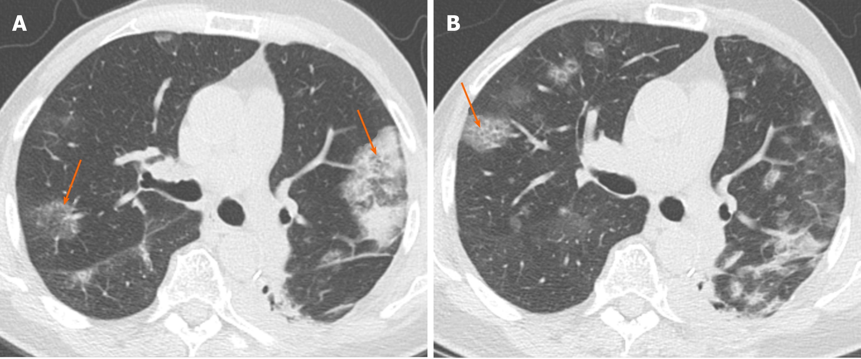
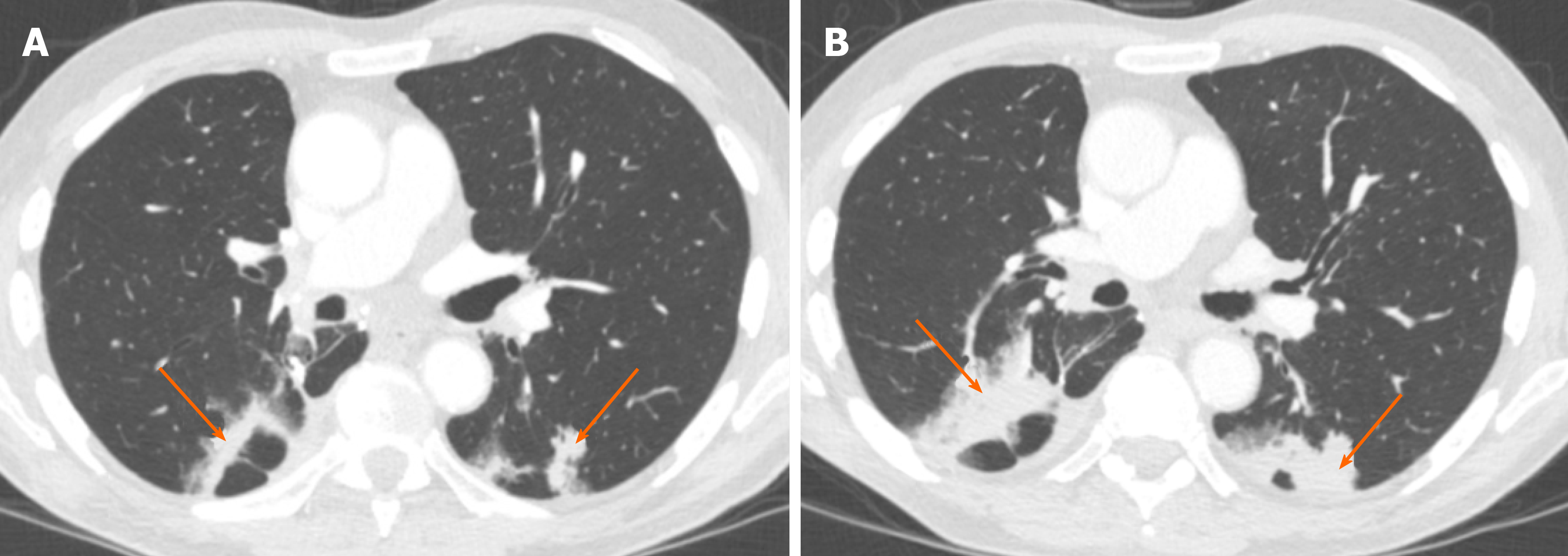
These small clustered branching and nodular opacities represent terminal airway mucous. These are due to filling of the. Tuberculosis manifesting as tree-in-bud opacities was made and the patient was started on anti-tuberculous treatment.
The purpose of this. Tree-in-bud TIB opacities are a common imaging finding on thoracic CT scan. However to our knowledge the relative frequencies of the causes have not been evaluated.
Find read and. Multiple causes for tree-in-bud TIB opacities an imaging pattern usually seen on chest CT have been reported. The patient died 15 days af-ter CT was performed.
This pattern is often seen in patients. TIB opacities represent a normally invisible branches of the bronchiole tree 1 mm in diameter that are severely impacted with mucous pus or fluid with resultant dilatation and. In clinical practice however it can reflect a.
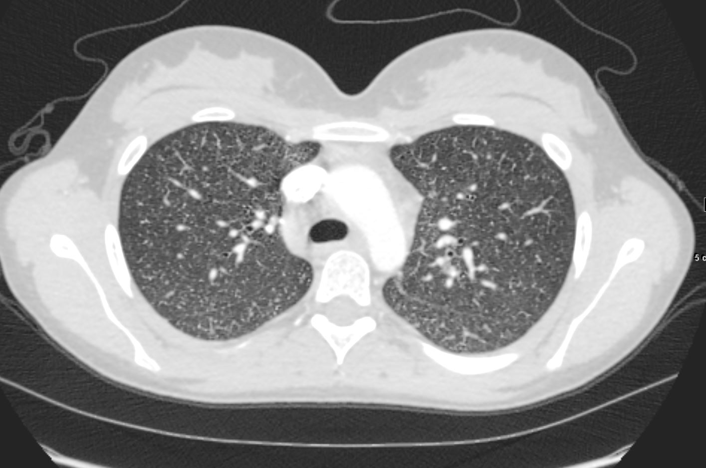
78 indicating the absenceresolution. INTRODUCTION Tree-in-bud TIB opacities are a subset of centrilobular nodules. What is tree-in-bud on CT scan.
TIB opacities represent a normally invisible branches of the bronchiole tree 1 mm in diameter that are severely impacted with mucous pus or fluid with resultant dilatation and. Treatment would include injection laryngoplasty and vocal cord medialisation9 Substantial numbers of patients with vocal cord palsy may develop malignant aetiologies later and. The tree-in-bud pattern is classically associated with endobronchial spread of tuberculosis or atypical mycobacterial infection.
After 2 months of treatment the cough and fever had subsided her. Tree-in-bud opacities appear as tiny centrilobular branching structures on CT most often in the lung periphery which resemble budding trees Figure 18-4.

Epos Trade

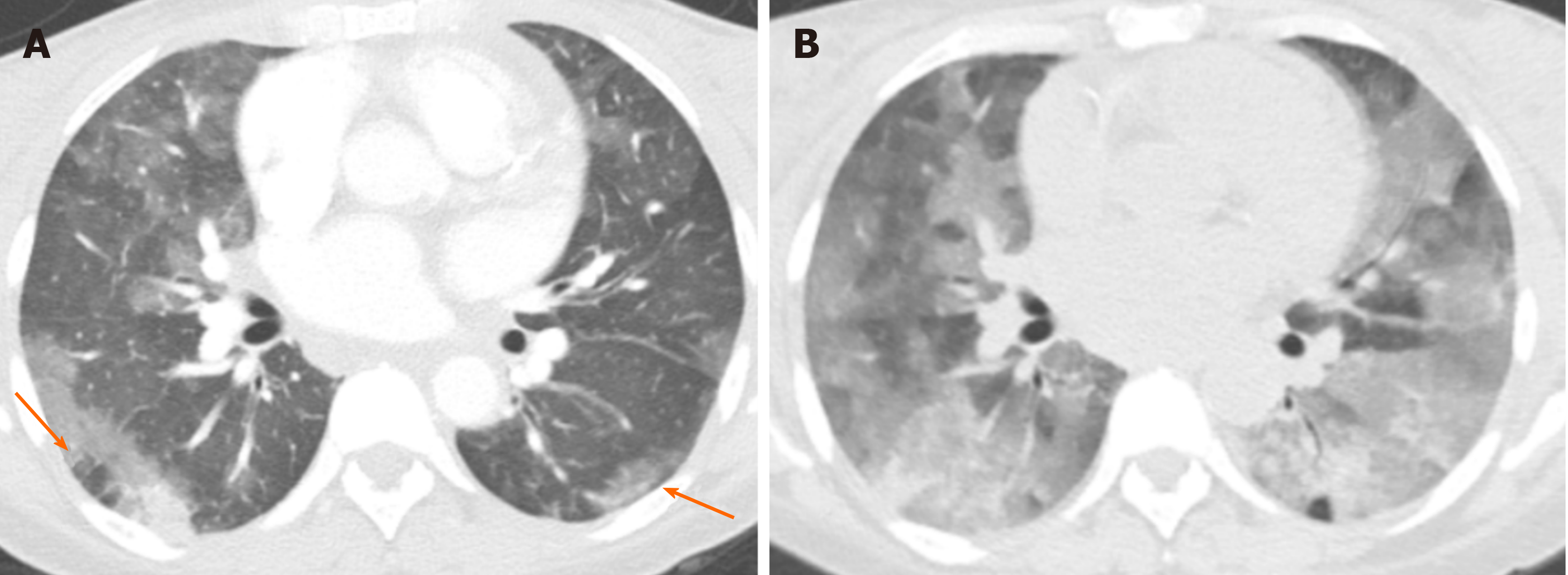
Review Of The Chest Ct Differential Diagnosis Of Ground Glass Opacities In The Covid Era Abstract Europe Pmc

Immune Related Adverse Events Associated With Checkpoint Inhibition In The Setting Of Car T Cell Therapy A Case Series Clinical Lymphoma Myeloma And Leukemia

Chest Ct Scan Condensation Areas Of Air Space Groundglass Opacities Download Scientific Diagram

Review Of The Chest Ct Differential Diagnosis Of Ground Glass Opacities In The Covid Era Abstract Europe Pmc
007lu Acute Histoplasmosis Lungs

Review Of The Chest Ct Differential Diagnosis Of Ground Glass Opacities In The Covid Era Abstract Europe Pmc
2

Review Of The Chest Ct Differential Diagnosis Of Ground Glass Opacities In The Covid Era Abstract Europe Pmc
Chronic Airspace Disease Review Of The Causes And Key Computed Tomography Findings
2
Chronic Airspace Disease Review Of The Causes And Key Computed Tomography Findings

Figure 3 From Tree In Bud Pattern Semantic Scholar

Scielo Brasil Diagnostic Performance Of The Rsna Proposed Classification For Covid 19 Pneumonia Versus Pre Pandemic Controls Diagnostic Performance Of The Rsna Proposed Classification For Covid 19 Pneumonia Versus Pre Pandemic Controls

Interstitial Lung Disease Ild Usual Interstitial Lung Disease Uip Lungs
Chronic Airspace Disease Review Of The Causes And Key Computed Tomography Findings
2
Chronic Airspace Disease Review Of The Causes And Key Computed Tomography Findings
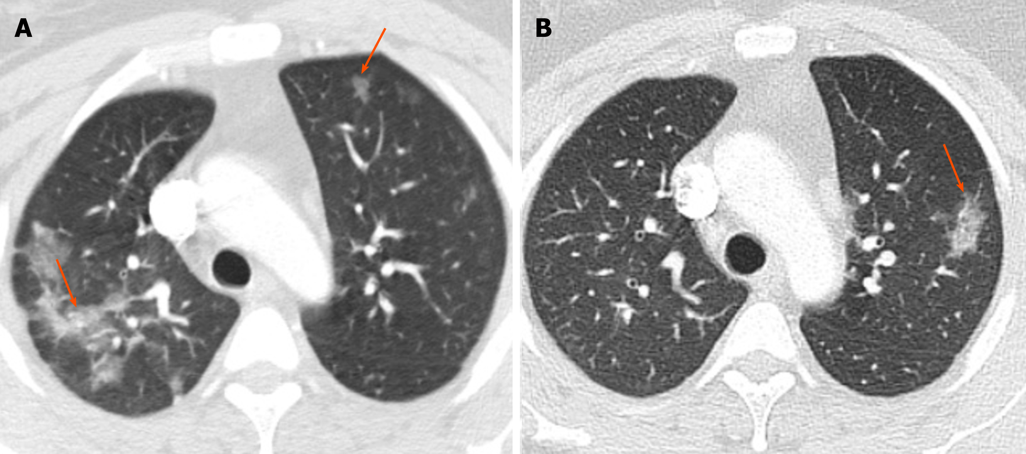
Chronic Airspace Disease Review Of The Causes And Key Computed Tomography Findings